How Do You Know if You Have Viral Conjunctivitis
Standing Pedagogy Activity
Conjunctivitis, also known as "pink eye", is inflammation of the conjunctiva. The three nearly common causes of conjunctivitis are viral, allergic, and bacterial, and the majority of cases are acquired by adenovirus. Conjunctivitis causes the middle to announced erythematous secondary to the dilation of blood vessels and is usually accompanied by increased vehement and/or mucoid discharge. This activity describes the risk factors, evaluation, and management of viral conjunctivitis and highlights the role of the interprofessional team in enhancing care delivery to affected patients.
Objectives:
-
Draw the common etiologies of viral conjunctivitis.
-
Distinguish viral causes of conjunctivitis from bacterial causes of conjunctivitis.
-
Identify the management strategies for viral conjunctivitis.
-
Explicate the importance of improving care coordination, with particular accent on communication between interprofessional medical teams, to enhance prompt and thorough delivery of care to patients with viral conjunctivitis.
Access free multiple choice questions on this topic.
Introduction
Conjunctivitis is one of the about common causes of red-eye and affects patients of all ages and socioeconomic class. Viral conjunctivitis is responsible for the majority of infectious conjunctivitis, accounting for up to 75% of cases. [1][ii] Characteristics of viral conjunctivitis include redness, blood vessel engorgement, ocular belch, hurting, photophobia, and pseudomembranes. At that place is a considerable economic and societal impact due to the costs of visits to the emergency department or general practitioner, diagnostic tests, prescription treatment, and time lost from work or school. Prescribing antibiotics in cases of viral conjunctivitis is one of the major costs of any healthcare system. The rates of antibiotic handling in the community for patients with infectious conjunctivitis in the United Kingdom range between 80% and 95%.[3][4] Improvement in diagnostic rates of viral conjunctivitis is estimated to have reduced inappropriate antibody prescribing and saved Usa $430 meg per year in the Usa.[ii] Specific treatments for viral conjunctivitis are currently being assessed in clinical trials. Due to the not-specificity of signs and symptoms, a thorough medical and ophthalmic history with clinical test should be obtained, especially in patients with atypical signs and a chronic course.
Etiology
The conjunctiva is a sparse semitransparent membrane that covers the white role of the eye called the sclera. The conjunctiva starts at the limbus of the cornea and covers both the sclera and posterior surface of the eyelids. The portion roofing the scleral is referred to every bit the bulbar conjunctiva, and the portion on the posterior surface of the lids is the palpebral conjunctiva.
The almost common crusade of viral conjunctivitis is adenoviruses. The adenovirus is part of the Adenoviridae family that consists of a nonenveloped, double-stranded DNA virus. Frequently associated infections caused by the adenovirus include upper respiratory tract infections, eye infections, and diarrhea in children. Children are nearly susceptible to viral infections, and adults tend to get more bacterial infections. Viral conjunctivitis can be obtained by straight contact with the virus, airborne transmission, and reservoir such as swimming pools. [5][six]
Most cases of viral conjunctivitis are highly contagious for 10-14 days. Washing hands and avoidance of eye contact are central to preventing manual to others.
Adenoviral Conjunctivitis
Upward to 90% of viral conjunctivitis cases are caused by adenoviruses.[7] In children pharyngoconjunctival fever (PCF) due to HAdV types 3, 4, and vii results in acute follicular conjunctivitis with fever, pharyngitis, periauricular lymphadenopathy. Epidemic keratoconjunctivitis (EKC) is the well-nigh severe ocular infection acquired by adenovirus and is classically associated with serotypes 8, 19, and 37. The cornea can exist afflicted by the viral replication in the epithelium and anterior stroma leading to superficial punctate keratopathy and subepithelial infiltrates.[8] Monotherapy with povidone-iodine ii% have demonstrated the resolution of symptoms.[ix] Further combinations of povidone-iodine with corticosteroids are undergoing phase 3 randomized controlled studies. Visual symptoms caused by subepithelial infiltrates tin can be debilitating and the use of tacrolimus, one% and two% cyclosporine A eye drops accept been shown to effective.[10][eleven][12]
Herpetic Conjunctivitis
Canker conjunctivitis is common in adults and children and associated with follicular conjunctivitis. Herpes simplex virus (HSV) is estimated to be responsible for 1.3 - iv.eight% of acute conjunctivitis cases.[xiii] Treatment with topical antiviral agents is aimed at reducing virus shedding and the development of keratitis. Varicella-zoster tin can cause conjunctivitis either by direct contact of the eye or skin lesions or inhalation of infected aerosolized particles, particularly with the involvement of the first and 2d branches of the trigeminal nerve.
Astute Hemorrhagic Conjunctivitis (AHC)
This is a very contagious class of viral conjunctivitis and symptoms include strange body sensation, epiphora, eyelid edema, conjunctival chemosis, and subconjunctival hemorrhage. A pocket-size proportion of patients experience systemic symptoms of fever, fatigue, limb hurting. Transmission is primarily via hand to eye contact and fomites.[14] Picornaviruses EV70 and coxsackievirus A24 variant (CA24v) are thought to be the responsible pathogens.[xv]
COVID-xix Conjunctivitis
The most recent isolated strain of coronavirus, COVID-19 has been reported to crusade conjunctivitis along with fever, coughing, respiratory distress, and death.[16] Retrospective and prospective studies testify that 1% to half dozen% of patients brandish COVID-19 related conjunctivitis, with conjunctival swabs being positive in ii.5% of cases.[17] Transmission through ocular tissues is incompletely understood. Ophthalmologists are at higher run a risk for COVID-19 infection due to the shut proximity to patients, equipment intense clinics, direct contact with patients' conjunctival mucosal surfaces, and high-volume clinics. Systemic COVID-19 is contracted through direct or airborne inhalation of respiratory droplets. Additional protective measures such every bit social distancing, wearing masks, reduced clinic volumes, and sterilization of surfaces should exist skillful to reduce transmission risk.
Epidemiology
Conjunctivitis, whether bacterial or viral, is a common problem that affects millions of Americans each year. In the United states of america, it is estimated that 1% of visits of a primary care physicians are related to conjunctivitis. While viral conjunctivitis is the most common cause, bacterial conjunctivitis is the 2d well-nigh common cause and distinguishing the two tin be a claiming for main care physicians. Often antibiotics are prescribed without practiced indication which could present an unnecessary financial burden on the patient and increase drug-resistant bacteria. Employers and schools typically crave those with conjunctivitis to remain out of their locations until the infection clears, potentially adding to the economical burden placed on those infected.[18]
Pathophysiology
Regardless of the etiology, most cases of conjunctivitis can be categorized as either papillary or follicular. Neither classification is pathognomonic for a particular disease entity. Papillary conjunctivitis produces a cobblestone arrangement of flattened nodules with central vascular cores. It is most commonly associated with an allergic immune response or is a response to a foreign body. Contained of the etiology, the histologic advent of papillary conjunctivitis is the aforementioned: closely packed, flat-topped projections, with numerous eosinophils, lymphocytes, plasma cells, and mast cells in the stroma surrounding a key vascular aqueduct.
Follicular conjunctivitis is seen in a variety of weather condition, including inflammation caused by pathogens such as viruses, bacteria, toxins, and topical medications. In contrast to papillae, follicles are small, dome-shaped nodules without a prominent central vessel. Histologically, a lymphoid follicle is situated in the subepithelial region and consists of a germinal heartwith young, proliferating lymphocytes surrounded by a ringof mature lymphocytes and plasma cells. The follicles in follicular conjunctivitis are typically most prominent in the inferior palpebral and forniceal conjunctiva.[nineteen]
History and Physical
The diagnosis of viral conjunctivitis is based on clinical and laboratory signs. Correct and early identification of etiology allows appropriate treatment and the abstention of longer-term complications.
Patients with viral conjunctivitis present with sudden onset strange body sensation, red optics, itching, lite sensitivity, burning, and watery discharge. Whereas with bacterial conjunctivitis, patients nowadays with all the above symptoms, but with mucopurulent discharge and mattering of the eyelids upon waking. Those presenting with viral conjunctivitis commonly have a recent history of upper respiratory tract infection or recent contact with a sick individual. Visual acuity is ordinarily at or nigh their baseline vision. The cornea can have subepithelial infiltrates that can decrease the vision and cause calorie-free sensitivity. The conjunctiva is injected (carmine) and tin too exist edematous. In some cases, a membrane or pseudomembrane can be appreciated in the tarsal conjunctiva. These are sheets of fibrin-rich exudates that are devoid of blood or lymphatic vessels. True membranes can pb to the development of subepithelial fibrosis and symblepharon, and as well bleed heavily on removal.[20] Follicles, small-scale, dome-shaped nodules without a prominent central vessel, can be seen on the palpebral conjunctiva. The majority of viral conjunctivitis patients will have follicles nowadays, only the presence of papillae does not rule out a viral etiology.[21] Palpation of the preauricular lymph nodes may reveal a reactive lymph node that is tender to the touch and will help differentiate viral conjunctivitis versus bacterial. With HSV, vesicles may appear on the face or eyelids and vision may exist affected. Corneal involvement may occur. With herpes zoster, at that place is a linear dermatomal pattern of vesicles. The conjunctiva is often red with a mucopurulent discharge.
Fever, angst, fatigue, presence of enlarged lymph nodes, and other constitutional signs help to differentiate viral conjunctivitis from other causes. Anisocoria and photophobia are associated with serious eye conditions including anterior uveitis, keratitis, and scleritis.[22] Systemic diseases associated with conjunctivitis include skin and mucous membrane diseases (acne rosacea, ichthyosis, xeroderma pigmentosum), collagen vascular diseases (systemic lupus erythematosus, rheumatoid arthritis, Sjogren's affliction, granulomatosis with polyangiitis), and autoimmunity (graft versus host disease, Steven-Johnson syndrome, and ocular cicatricial pemphigoid).
Evaluation
Laboratory testing is typically not indicated unless the symptoms are non resolving and infection last longer than iv weeks. Laboratory testing tin can be indicated in certain situations such as a suspected chlamydial infection in a newborn, an immune-compromised patient, excessive amounts of discharge, or suspected gonorrhea co-infection. In the office, physicians can run tests to positively place adenovirus with a specificity and sensitivity of 89% and 94%, respectively. However, ophthalmologists commonly can make the diagnosis clinically with confirmational additional testing.[23]
Regarding adenovirus testing, the gold standard has traditionally been cell culture because once the virus is isolated, the diagnosis is definitive and the label of the virus tin can be undertaken.[24] Disadvantages include cost and increased time associated with cell culture-based testing. The mainstay of viral conjunctivitis testing in the developed earth is the detection of viral DNA by polymerase concatenation reaction (PCR). PCR for adenovirus has been shown to exist 93% sensitive and 97.iii% specific from conjunctival swabs, with similar values, plant for HSV diagnosis.[24] A recent development has been the cosmos of a rapid detection testing platform for adenovirus, the AdenoPlus assay (Rapid Pathogen Screening Inc., Sarasota, Florida, Us). This is a swift office-based exam designed to detect 53 serotypes of adenovirus and provide a result within x minutes.[25] Studies accept demonstrated high specificity values of 92% to 98% in detecting adenoviral conjunctivitis. [26][27][28] However, the test has lower sensitivity reported compared with PCR analysis.[26][27][28] Rapid detection testing can be more useful in the time to come one time viral conjunctivitis treatments reach the clinical setting.[29]
Treatment / Direction
Treatment for viral conjunctivitis is aimed at symptomatic relief and non to eradicate the self-limiting viral infection. The resolution of conjunctivitis can take up to three weeks. Handling includes using artificial tears for lubrication four times a day or up to ten times a day with preservative-free tears. Cool compresses with a wet washcloth to the periocular area may provide symptomatic relief. Preventing the spread of infection to the other center or other people requires the patient to practice skillful paw hygiene with frequent washing, avoidance of sharing towels or linens, and fugitive touching their eyes. A person is thought to shed the virus while their eyes are cerise and tearing.
If a membrane or pseudomembrane is present, it tin be peeled at the slit lamp to improve patient condolement and prevent any scar formation from occurring. These membranes can either be peeled with a jeweler forceps or a cotton fiber swab soaked with topical anesthetic. Topical steroids can aid with the resolution of symptoms. However, they can also crusade the shedding of the virus to last longer. Patients should be informed that they are highly contagious and should refrain from work or schoolhouse until their symptoms resolve. While using steroids, they may still shed the virus without the visual symptoms that would indicate that they have an infection. Steroids should be reserved for patients with decreased vision due to their subepithelial infiltrates or astringent conjunctival injection causing more than the expected discomfort.[30][31]
The use of povidone-iodine, a non-specific disinfectant, is a promising new treatment for adenoviral conjunctivitis.[25] This is an inexpensive and widely bachelor antiseptic solution which is used as part of the aseptic preparation for ocular surgery. It is able to impale extracellular organisms but has no effect intracellularly. It does non induce drug resistance because its machinery of action is not immunologically dependent. A single dose of 2.v% povidone-iodine in infants with adenoviral conjunctivitis resulted in a reduction in symptom severity and reduced recovery time without meaning side effects.[32]
Topical corticosteroids solitary are contraindicated in epithelial herpes simplex keratitis and are associated with prolonged viral shedding and infection.[29] When used in combination with an anti-infective, corticosteroids have demonstrated good tolerability and efficacy in treating the inflammatory and infectious components of conjunctivitis.[33][34]
Differential Diagnosis
While the most mutual causes of conjunctivitis are viral or bacterial and due to allergic reactions, there are other causes of conjunctivitis that should be considered when treatment does non improve symptoms. Uveitis is a local autoimmune reaction that causes the middle to become inflamed and is commonly mistaken for conjunctivitis. Uveitis can be a local reaction that is idiopathic or a manifestation of a systemic autoimmune disease such as rheumatoid arthritis, lupus, or ankylosis spondylitis. Systemic autoimmune affliction such Sjogren syndrome or Stevens-Johnson syndrome tin can also mimic conjunctivitis by presenting with conjunctival erythema and discharge.
A systemic workup should be initiated if whatever of these diseases are suspected. The most life or sight-threatening masqueraders of conjunctivitis include clangorous carotid fistula, orbital cellulitis, and orbital hemorrhage. In cavernous carotid fistulas, an abnormal communication between the arterial and venous circulation form causing vasodilation of the venous arrangement. Afterward, the fistula can cause rupture and hemorrhage, leading to irreversible harm to the eye and/or death. Due to venostasis, the ophthalmic vein dilates and causes congestion of the episcleral vessels mimicking conjunctivitis. Proptosis and a pulsatile earth can help differentiate between a fistula and conjunctivitis. Orbital cellulitis is an infection posterior to the septum and involves the orbital contents. Patients present with similar symptoms of conjunctivitis, merely will also take pain with eye movements or fifty-fifty restricted eye movements. Lastly, an orbital hemorrhage is an ophthalmic emergency. The crusade is mostly traumatic, simply it is possible to have a spontaneous hemorrhage, specially in patients on anticoagulants, that nowadays with proptosis, tight eyelids, and erythema of the conjunctiva.
Prognosis
The majority of cases of virus conjunctivitis resolve on their own. In rare cases, chronic infection may occur. Near cases resolve inside 14-xxx days. In some patients, photophobia, diminished vision and glare may be a problem.
Complications
-
Punctate keratitis
-
Bacterial superinfection
-
Conjunctival scarring
-
Corneal ulceration
-
Chronic infection
Deterrence and Patient Education
The vast majority of viral conjunctivitis is acquired by adenoviruses, and frequent hand washing, disinfection, and isolation of conjunctivitis patients can reduce further community transmission. In that location is no unmarried treatment modality for viral conjunctivitis. Symptom relief with artificial tears, cold-compresses, and antihistamines tin be effective. Antibiotic drops tin potentially increase bacterial resistance and membranes or pseudomembrane require removal to reduce discomfort and scarring. The use of topical corticosteroids is indicated in select cases in the presence of membrane germination and subepithelial infiltration associated with photophobia and decreased vision.
Enhancing Healthcare Team Outcomes
The majority of patients with viral conjunctivitis are first managed by the primary care giver, pediatrician or nurse practitioner. Fifty-fifty though viral conjunctivitis is a benign condition, information technology is contagious tin can be easily transmitted to others. The key is patient education.
Patients need to be educated that the condition is harmless and will resolve spontaneously. Hand washing should exist emphasized since the infection is highly contagious. Parents and teachers should be educated on the importance of isolation in schoolhouse to prevent epidemics. All patients with viral conjunctivitis who wear contact lenses should be told to refrain from wearing them until the symptoms have subsided.
The emergency section should take a special room for patients presenting with conjunctivitis to preclude spread to other patients. signs should exist placed on the doors about not shaking hands and washing hands at every opportunity. One time the patient has been seen, he or she must be individually escorted out of the clinic to avert contact with other patients. Many lawsuits take resulted in the past after patients in the emergency room acquired conjunctivitis from infected individuals, sitting in the same location.
While long-term sequelae are rare, chronic viral conjunctivitis can atomic number 82 to a poor quality of life. Most cases take ane-iv weeks to recover without handling. The morbidity is rare simply tin include corneal ulceration and punctate keratitis. Follow upward with an ophthalmologist is recommended to ensure that no complications have occurred.[35][36] (Level 5)
Review Questions

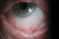
Figure
Viral conjunctivitis. Image courtesy S Bhimji
Figure
Atypical features such every bit chronic symptoms, relapsing/remitting course, and signs such as symblepharon, loss of the plica, shortening of conjunctival fornices propose a non-viral crusade of conjunctivitis. Contributed by Dr Lanxing Fu.
References
- 1.
-
Groovy M, Thompson M. Handling of Acute Conjunctivitis in the United states of america and Bear witness of Antibiotic Overuse: Isolated Issue or a Systematic Trouble? Ophthalmology. 2017 Aug;124(8):1096-1098. [PubMed: 28734327]
- 2.
-
Udeh BL, Schneider JE, Ohsfeldt RL. Toll effectiveness of a point-of-intendance examination for adenoviral conjunctivitis. Am J Med Sci. 2008 Sep;336(3):254-64. [PubMed: 18794621]
- 3.
-
Rietveld RP, ter Riet G, Bindels PJ, Schellevis FG, van Weert HC. Practice full general practitioners adhere to the guideline on infectious conjunctivitis? Results of the Second Dutch National Survey of General Practice. BMC Fam Pract. 2007 Sep sixteen;8:54. [PMC costless article: PMC2034564] [PubMed: 17868475]
- 4.
-
Everitt H, Piffling P. How do GPs diagnose and manage acute infective conjunctivitis? A GP survey. Fam Pract. 2002 December;19(6):658-60. [PubMed: 12429670]
- 5.
-
Li J, Lu X, Jiang B, Du Y, Yang Y, Qian H, Liu B, Lin C, Jia L, Chen L, Wang Q. Adenovirus-associated astute conjunctivitis in Beijing, China, 2011-2013. BMC Infect Dis. 2018 Mar 20;18(1):135. [PMC free commodity: PMC5859447] [PubMed: 29558885]
- half-dozen.
-
Sow Equally, Kane H, Ka AM, Hanne FT, Ndiaye JMM, Diagne JP, Nguer M, Sow Southward, Saheli Y, Sy EHM, De Meideros Quenum ME, Ndoye Roth PA, Ba EA, Ndiaye PA. [Senegalese experience with acute viral conjunctivitis]. J Fr Ophtalmol. 2017 Apr;twoscore(4):297-302. [PubMed: 28342559]
- 7.
-
Azari AA, Barney NP. Conjunctivitis: a systematic review of diagnosis and handling. JAMA. 2013 October 23;310(16):1721-9. [PMC costless article: PMC4049531] [PubMed: 24150468]
- 8.
-
Chigbu DI, Labib BA. Pathogenesis and direction of adenoviral keratoconjunctivitis. Infect Drug Resist. 2018;11:981-993. [PMC free article: PMC6054290] [PubMed: 30046247]
- nine.
-
Trinavarat A, Atchaneeyasakul LO. Treatment of epidemic keratoconjunctivitis with two% povidone-iodine: a pilot report. J Ocul Pharmacol Ther. 2012 Feb;28(one):53-8. [PubMed: 21916618]
- 10.
-
Levinger Due east, Slomovic A, Sansanayudh W, Bahar I, Slomovic AR. Topical treatment with 1% cyclosporine for subepithelial infiltrates secondary to adenoviral keratoconjunctivitis. Cornea. 2010 Jun;29(six):638-twoscore. [PubMed: 20458220]
- eleven.
-
Berisa Prado S, Riestra Ayora AC, Lisa Fernández C, Chacón Rodríguez M, Merayo-Lloves J, Alfonso Sánchez JF. Topical Tacrolimus for Corneal Subepithelial Infiltrates Secondary to Adenoviral Keratoconjunctivitis. Cornea. 2017 Sep;36(9):1102-1105. [PubMed: 28704319]
- 12.
-
Arici C, Mergen B. Late-term topical tacrolimus for subepithelial infiltrates resistant to topical steroids and ciclosporin secondary to adenoviral keratoconjunctivitis. Br J Ophthalmol. 2021 May;105(five):614-618. [PubMed: 32563992]
- 13.
-
Sheikh A, Hurwitz B, van Schayck CP, McLean S, Nurmatov U. Antibiotics versus placebo for acute bacterial conjunctivitis. Cochrane Database Syst Rev. 2012 Sep 12;(9):CD001211. [PubMed: 22972049]
- fourteen.
-
Langford MP, Anders EA, Burch MA. Acute hemorrhagic conjunctivitis: anti-coxsackievirus A24 variant secretory immunoglobulin A in astute and convalescent tear. Clin Ophthalmol. 2015;9:1665-73. [PMC costless article: PMC4574852] [PubMed: 26392747]
- 15.
-
Zhang Fifty, Zhao Due north, Huang X, Jin X, Geng X, Chan TC, Liu S. Molecular epidemiology of acute hemorrhagic conjunctivitis caused by coxsackie A type 24 variant in China, 2004-2014. Sci Rep. 2017 Mar 23;7:45202. [PMC complimentary article: PMC5362916] [PubMed: 28332617]
- 16.
-
Huang C, Wang Y, Li X, Ren L, Zhao J, Hu Y, Zhang L, Fan G, Xu J, Gu X, Cheng Z, Yu T, Xia J, Wei Y, Wu Westward, Xie X, Yin Westward, Li H, Liu M, Xiao Y, Gao H, Guo L, Xie J, Wang One thousand, Jiang R, Gao Z, Jin Q, Wang J, Cao B. Clinical features of patients infected with 2019 novel coronavirus in Wuhan, China. Lancet. 2020 Feb 15;395(10223):497-506. [PMC free article: PMC7159299] [PubMed: 31986264]
- 17.
-
Danesh-Meyer HV, McGhee CNJ. Implications of COVID-19 for Ophthalmologists. Am J Ophthalmol. 2021 Mar;223:108-118. [PMC gratis article: PMC7506460] [PubMed: 32976847]
- 18.
-
Binder AM, Biggs HM, Haynes AK, Chommanard C, Lu Ten, Erdman DD, Watson JT, Gerber SI. Human Adenovirus Surveillance - United States, 2003-2016. MMWR Morb Mortal Wkly Rep. 2017 Oct 06;66(39):1039-1042. [PMC gratis commodity: PMC5720882] [PubMed: 28981484]
- 19.
-
Alfonso SA, Fawley JD, Alexa Lu X. Conjunctivitis. Prim Care. 2015 Sep;42(three):325-45. [PubMed: 26319341]
- xx.
-
Chintakuntlawar AV, Chodosh J. Cellular and tissue architecture of conjunctival membranes in epidemic keratoconjunctivitis. Ocul Immunol Inflamm. 2010 Oct;eighteen(5):341-5. [PMC free article: PMC2974573] [PubMed: 20735288]
- 21.
-
Marinos E, Cabrera-Aguas M, Watson SL. Viral conjunctivitis: a retrospective report in an Australian hospital. Cont Lens Inductive Middle. 2019 Dec;42(6):679-684. [PubMed: 31300283]
- 22.
-
Narayana S, McGee Southward. Bedside Diagnosis of the 'Red Eye': A Systematic Review. Am J Med. 2015 Nov;128(11):1220-1224.e1. [PubMed: 26169885]
- 23.
-
Sethuraman U, Kamat D. The scarlet centre: evaluation and management. Clin Pediatr (Phila). 2009 Jul;48(6):588-600. [PubMed: 19357422]
- 24.
-
Elnifro EM, Cooper RJ, Klapper PE, Yeo AC, Tullo AB. Multiplex polymerase chain reaction for diagnosis of viral and chlamydial keratoconjunctivitis. Invest Ophthalmol Vis Sci. 2000 Jun;41(7):1818-22. [PubMed: 10845603]
- 25.
-
Kaufman HE. Adenovirus advances: new diagnostic and therapeutic options. Curr Opin Ophthalmol. 2011 Jul;22(4):290-three. [PubMed: 21537185]
- 26.
-
Kam KY, Ong HS, Bunce C, Ogunbowale L, Verma S. Sensitivity and specificity of the AdenoPlus point-of-care system in detecting adenovirus in conjunctivitis patients at an ophthalmic emergency section: a diagnostic accuracy study. Br J Ophthalmol. 2015 Sep;99(9):1186-nine. [PubMed: 25824258]
- 27.
-
Sambursky R, Trattler W, Tauber S, Starr C, Friedberg Chiliad, Boland T, McDonald Chiliad, DellaVecchia Chiliad, Luchs J. Sensitivity and specificity of the AdenoPlus test for diagnosing adenoviral conjunctivitis. JAMA Ophthalmol. 2013 Jan;131(1):17-22. [PubMed: 23307204]
- 28.
-
Holtz KK, Townsend KR, Furst JW, Myers JF, Binnicker MJ, Quigg SM, Maxson JA, Espy MJ. An Assessment of the AdenoPlus Point-of-Care Test for Diagnosing Adenoviral Conjunctivitis and Its Effect on Antibiotic Stewardship. Mayo Clin Proc Innov Qual Outcomes. 2017 Sep;1(2):170-175. [PMC complimentary commodity: PMC6135020] [PubMed: 30225413]
- 29.
-
Jhanji V, Chan TC, Li EY, Agarwal K, Vajpayee RB. Adenoviral keratoconjunctivitis. Surv Ophthalmol. 2015 Sep-Oct;60(5):435-43. [PubMed: 26077630]
- 30.
-
Usher P, Keefe J, Crock C, Chan E. Appropriate prescribing for viral conjunctivitis. Aust Fam Physician. 2014 November;43(eleven):748-ix. [PubMed: 25551873]
- 31.
-
Shiota H, Ohno S, Aoki Yard, Azumi A, Ishiko H, Inoue Y, Usui Northward, Uchio E, Kaneko H, Kumakura S, Tagawa Y, Tanifuji Y, Nakagawa H, Hinokuma R, Yamazaki South, Yokoi Due north. [Guideline for the nosocomial infections of adenovirus conjunctivitis]. Nippon Ganka Gakkai Zasshi. 2009 Jan;113(1):25-46. [PubMed: 19227929]
- 32.
-
Özen Tunay Z, Ozdemir O, Petricli IS. Povidone iodine in the handling of adenoviral conjunctivitis in infants. Cutan Ocul Toxicol. 2015 Mar;34(one):12-v. [PubMed: 24678746]
- 33.
-
Kovalyuk N, Kaiserman I, Mimouni M, Cohen O, Levartovsky S, Sherbany H, Mandelboim M. Handling of adenoviral keratoconjunctivitis with a combination of povidone-iodine 1.0% and dexamethasone 0.ane% drops: a clinical prospective controlled randomized report. Acta Ophthalmol. 2017 Dec;95(viii):e686-e692. [PubMed: 28342227]
- 34.
-
Faraldi F, Papa Five, Rasà D, Santoro D, Russo Due south. Netilmicin/dexamethasone fixed combination in the treatment of conjunctival inflammation. Clin Ophthalmol. 2013;7:1239-44. [PMC free article: PMC3699304] [PubMed: 23836952]
- 35.
-
Levine J, Snyder RW. Practical ophthalmic microbiology. J Ophthalmic Nurs Technol. 1999 Mar-Apr;18(ii):50-nine; quiz 74-5. [PubMed: 10409996]
- 36.
-
Uçakhan Ö, Yanik Ö. The Utilise of Cast Contact Lenses in Adenoviral Keratoconjunctivitis. Eye Contact Lens. 2016 Nov;42(6):388-391. [PubMed: 26671622]
Source: https://www.ncbi.nlm.nih.gov/books/NBK470271/
0 Response to "How Do You Know if You Have Viral Conjunctivitis"
Post a Comment